Muscles of the Torso
The torso, also called the trunk, is the central area of the body which contains over 40 bones and 30 pairs of skeletal muscles. It provides attachments points for the proximal ends of the upper and lower limbs, as well as the neck and head. The torso contains all of the organs or viscera of the body (except the brain). The primary functions of the muscles of the torso are to enable movement of the lumbar and thoracic spine, ribs and proximal limbs, to facilitate breathing, and protect the the vital organs.
What is the torso of the body? The torso is the central region of the body that connects the upper and lower extremities. Areas within the torso include the chest, abdomen, thoracic and lumbar. Many of the bones of the axial skeleton, such as the rib cage, sternum, and spine (thoracic, lumbar, sacral and coccyx) are within the torso. Muscles of the torso include the abdominals, pectorals, erector spinae, serratus anterior and posterior, intercostals, latissimus dorsi, and rhomboids.
This muscles of the torso review is designed to help massage therapy students learn muscles for school or for the Massage & Bodywork Licensing Exam (MBLEx). Exam candidates need to know the origins, insertions and actions (OIA) for all skeletal muscles of the body. You may encounter questions about muscle OIA in the kinesiology or anatomy content areas of the massage exam. The muscle tables below also include the muscle innervations which are helpful to know. This reference guide also covers muscle groups of the anterior and posterior torso.
Muscles of the torso for massage therapists
Muscles and the connective tissues that surround them are what most massage therapists and bodyworkers target with their manual therapy techniques. One of the most common complaints and reasons that clients seek massage therapy is to relieve their back pain. While back pain can have numerous causes, including everything from kidney stones, herniated discs and fibromyalgia to overuse injuries, back pain is commonly associated with increased muscle tension (hypertonicity) of the back muscles. Massage therapists who have a good understanding of anatomy and know how to correctly apply therapeutic techniques will have a better chance of relieving their client’s back pain.
It is important for all manual therapists to understand the locations, layers, origins, insertions, actions and fiber directions of the muscles that they work on. This knowledge is particularly important when providing therapeutic treatments to address a specific client concern. For example, when you are trying to target the belly of a muscle to relieve a trigger point.
Massage therapists who are preparing to take the MBLEx exam can expect to encounter several questions about muscle attachment sites, actions, or muscle position relative to other structures of the body. As stated by the FSMTB, ‘you won’t be asked to name the origin, insertion and action of every muscle’. But since you don’t know which muscles they will ask about, you do need to know the OIA of every skeletal muscle that a massage therapist would normally work on.
***MBLEx study tip: combining the study of written materials and hands-on practice with a massage study buddy is a great way to learn muscles. Here is a simple 5-step process to study 1 muscle at a time:
(1) Briefly review a muscle’s origin, insertion and action
(2) Trace the outline of the muscle on your study partner, palpating the origin and insertion
(3) Demonstrating the action(s) of the muscle
(4) Quiz each other on the muscle’s OIA
(5) Review the OIA on the muscle table
When studying the muscles of the torso, massage therapists should also review the locations of these muscles relative to the areas of caution for massage, also called massage endangerment sites. Massage areas of caution on the anterior torso include the anterior pectoral and deltoid region, breast tissue, xiphoid process of the sternum, the lower ribs, umbilicus, and the urinary bladder and pubic regions. Endangerment sites of the posterior torso include the lower ribs, the kidney area, and the coccyx.


Overview of torso muscles and groups
The torso region of the body is often divided into subregions when discussing the structures and functions of the area. It can be divided into the upper torso, also called thorax, and the lower torso, which includes the abdomen and pelvic areas. The torso can also be divided into the anterior and posterior regions.
There is some overlap between what is considered the torso and what is considered the lower extremity. The pelvic and hip areas may be included in discussion of the torso or the lower extremity. Likewise, some of the muscles around the scapula may be covered when discussing the torso or the muscles of the upper extremity.
There are several major muscle groups of the torso. The main way that muscles of the torso are grouped is either by their location or by their function.
Core muscles support, move and stabilize the trunk
The core of the body refers to the central or core region of the body, which lies within the torso, especially the lower and middle regions of the torso. Muscles of the core include muscles of the anterior and posterior torso, and the pelvis.
- Abdominal muscle group (transversus abdominis, internal and external obliques, rectus abdominis)
- Middle and lower sections of the erector spinae group (spinalis, longissimus, iliocostalis)
- Multifidus, quadratus lumborum, diaphragm, psoas
- Minor (peripheral) core muscles include: trapezius, latissimus dorsi and gluteus maximus.
The main functions of core muscles include stabilizing structures of the thoracic and lumbopelvic regions (e.g. spine, joints, organs), supporting posture especially when moving, standing, or sitting, coordinating movement and transferring force between the upper and lower body. There’s a lot of mechanical stress at the core region because it is where the upper body intersects with the lower body. This is why it is important to keep the core muscles strong. Using good body mechanics is another way to reduce mechanical stress and prevent injury to this region.
Muscle groups of the anterior and lateral torso
- Pectorals or chest. Pectoralis major and pectoralis minor.
- Abdominals (from superficial to deep): rectus abdominis, external obliques, internal obliques, transversus abdominis.
Muscle groups of the posterior torso
- Erector spinae group. Spinalis, longissimus, iliocostalis. (From medial to lateral: SLI)
- Transversospinalis group. Semispinalis, multifidus, rotatores, interspinales, intertransversarii.

Learn Muscles for Massage Therapy
The Learn Muscles in 60 Days online course is designed to help massage therapy students learn and remember the muscles, origins, insertions and actions for school and for the MBLEx. Check your comprehension and retention with lesson quizzes and longer section quizzes. This muscle course is also beneficial for other health and wellness professionals who need to master their knowledge of the muscles of the body.
Torso muscles grouped by function
These muscle groups function together to perform specific tasks or movements, regardless of where they are located on the torso.
- Primary breathing muscles. Diaphragm (primarily), external intercostals. These muscles contract to enlarge the thoracic cavity and draw air in. During relaxed exhalation, air is expelled when these muscles relax due to the elastic recoil of the lungs and the surrounding muscles.
- Accessory breathing muscles. These are divided into accessory inspiratory muscles (breathing in) which expand the chest cavity to bring in more air. And accessory expiratory muscles (breathing out), which forcefully compress the thoracic and abdominal cavities to expel air from the lungs. Accessory breathing muscles are recruited to assist the diaphragm when more air exchange is needed, like when exercising or when in respiratory distress.
- Accessory muscle of inspiration. Sternocleidomastoid (SCM), scalenes (anterior, middle and posterior), external intercostals, serratus posterior superior and inferior, pectoralis major and minor, trapezius, latissimus dorsi, and quadratus lumborum.
- Accessory muscles of expiration. Internal and innermost intercostals, all 4 muscle layers of the abdominal muscle group (rectus abdominis, external and internal obliques, and transversus abdominis).
- Scapula muscles. Also called the periscapular muscles, these muscles move the scapula to assist with movement of the upper extremity. Scapular muscles are located at the posterior, lateral and anterior aspects of the torso.
- Elevators of the scapula. Upper trapezius, levator scapula.
- Depressors of the scapula. Lower trapezius, pectoralis minor.
- Protractors of the scapula. Serratus anterior, pectoralis minor.
- Retractors of the scapula. Rhomboid, middle trapezius.
- Upward rotators of the scapula. Upper and lower trapezius, serratus anterior. (Upward rotation of the scapula pivots the glenoid fossa upward).
- Downward rotators of the scapula. Levator scapula, rhomboids, pectoralis minor. (Downward rotation of the scapula pivots the glenoid fossa downward).
- Pelvic floor muscles. The pelvic floor is a dome-shaped muscular structure between the coccyx and pubic bone, which separates the pelvic cavity from the perineal region. It is composed of numerous ligaments and small muscles. You are unlikely to see specific questions about pelvic floor muscles on the MBLEx. The pelvic floor plays an important role in normal bodily functions such as maintaining continence, voiding, and childbirth.

Study with our complete MBLEx Prep Course!
Our comprehensive MBLEx Prep Course provides massage students with a thorough review of all 7 content areas of the FSMTB Massage & Bodywork Licensing Exam. Learn muscle anatomy and kinesiology, physiology, pathology, client assessment, ethics, and professional practice guidelines. Includes practice exams and quizzes.
Muscle OIA of the Anterior Torso
| Muscle | Origin | Insertion | Action | Innervation |
|---|---|---|---|---|
| Diaphragm | Internal surfaces of the lower 6 ribs and costal cartilages, xiphoid process of the sternum, and anterior surfaces of lumbar vertebrae 1-3. | Central tendon of the diaphragm | Compresses the abdominal viscera inferiorly which increases the volume of the thoracic cavity (inspiration) | Phrenic nerve (C3-5) |
| External intercostal | Inferior border of ribs 1-11 | Superior border of ribs 2-12 directly below the rib of origin | Elevates ribs during inspiration (breathing in). Also maintains intercostal spaces and supports rib cage. | Intercostal nerves |
| Innermost intercostal | Inferior border of each rib (costal groove) | Superior border of each rib immediately below origin | Depresses ribs during forced expiration (breathing out). Also maintains intercostal spaces and supports rib cage. | Intercostal nerves |
| Internal intercostal | Superior border of ribs 2-12 | Inferior border of ribs 1-11 immediately above the rib of origin | Depresses ribs during forced expiration (breathing out). Also maintains intercostal spaces and supports rib cage. | Intercostal nerves |
| External oblique (abdominal) | External surfaces of lower 8 ribs (ribs 5-12) | Linea alba, abdominal aponeurosis, pubic crest & tubercle, anterior half of iliac crest | Bilateral contraction: flexion of trunk, compression of abdominal contents. Unilateral contraction: lateral flexion of trunk (sidebending). Rotation of trunk to opposite side. | Intercostal nerves (T7-11). Also subcostal, iliohypogastric and ilioinguinal nerves. |
| Internal oblique (abdominal) | Thoracolumbar fascia, anterior iliac crest, lateral inguinal ligament | Costal cartilages of lower 3 ribs, linea alba, abdominal aponeurosis and pubic crest | Bilateral contraction: flexion of trunk, compression of abdominal contents. Unilateral contraction: lateral flexion of trunk (sidebending). Rotation of trunk to same side. | Intercostal nerves (T7-11). Also subcostal, iliohypogastric and ilioinguinal nerves. |
| Pectoralis major | Clavicular head: medial half of clavicle (anterior surface) Sternal head: anterior surface of sternum and costal cartilages of ribs 1-6 | Greater tubercle and lateral lip of bicipital groove of humerus | Flexion, adduction, horizontal adduction, and medial rotation of humerus at the glenohumeral joint. | Medial and lateral pectoral nerves (C5-T1) |
| Pectoralis minor | Anterior surface of costal cartilages and ribs 3-5 | Coracoid process of scapula (medial aspect) | Protraction, depression and downward rotation of the scapula at the scapulothoracic joint. | Medial pectoral nerve (C8,T1) |
| Rectus abdominis | Pubic symphysis and crest | Xiphoid process of sternum, and inferior surfaces of costal cartilages of ribs 5-7 | Flexion of trunk and compression of abdominal viscera. | Intercostal nerves (T7-12) |
| Serratus anterior | Superior and lateral surfaces of upper 8 or 9 ribs (at sides of rib cage) | Anterior surface of superior angle, medial border and inferior angle of scapula | Protraction and upward rotation of the scapula. Stabilizes scapula against the thoracic wall. | Long thoracic nerve (C5-C7) |
| Subclavius | Sternal end of first rib and costal cartilage | Inferior surface of clavicle (middle third) | Depression of clavicle at the sternoclavicular (SC) joint | Nerve to subclavius (C5,6) |
| Transversus abdominis | Internal surfaces of costal cartilages 7-12. Thoracolumbar fascia. Anterior part of iliac crest. Lateral part of inguinal ligament | Abdominal aponeurosis, linea alba, and pubic crest. | Compressesion of abdomen | Intercostal nerves (T7-11). Also subcostal, iliohypogastric and ilioinguinal nerves. |

Muscle Flashcards
All of our memberships (Gold, Silver and Bronze) now include 115 muscle flashcards to help students learn muscle origins, insertions, actions and innervations for massage school or for the MBLEx.
Muscle OIA of the Posterior Torso
| Muscle | Origin | Insertion | Action | Innervation |
|---|---|---|---|---|
| Iliocostalis cervicis | Superior borders of posterior ribs 3-6 | Transverse processes of C4-6 | Bilateral contraction: extension of cervical spine Unilateral contraction: lateral flexion of cervical spine to same side | Posterior rami of cervical and thoracic spinal nerves |
| Iliocostalis thoracic | Superior borders of lower 7 ribs, medial to the angle of ribs | Angles of ribs 1-6, transverse process of C7 | Bilateral contraction: extension of thoracic vertebral column Unilateral contraction: lateral flexion of thoracic spine to same side | Posterior rami of thoracic spinal nerves |
| Iliocostalis lumborum | Sacrum, iliac crest, and spinous processes of T11 – L5 vertebrae. | Inferior borders of lower 7 ribs | Bilateral contraction: extension of lumbar vertebral column, depresses ribs Unilateral contraction: lateral flexion of lumbar spine to same side | Posterior rami of lumbar spinal nerves |
| Interspinales | Spinous processes of each vertebrae | Spinous process of adjacent vertebra (immediately above) | Extension of spine, especially in the cervical and thoracic regions | Spinal nerves |
| Intertransversarii | Upper border of transverse process | Lower border of transverse process of adjacent vertebra (immediately above) | Lateral flexion of the spine, and stabilizes the spine. | Spinal nerves |
| Latissimus dorsi | Spinous processes of T7-L5 vertebrae, sacrum, thoracolumbar fascia, posterior iliac crest, and lower 3 or 4 ribs | Bicipital (intertubercular) groove of humerus | Extension, adduction and medial rotation of humerus at glenohumeral joint | Thoracodorsal nerve (C7,8) |
| Longissimus capitis | Transverse processes of T1-5 and articular processes of C4-7 | Posterior part of mastoid process of the temporal bone. | Bilateral contraction: extension of neck Unilateral contraction: lateral cervical flexion, and rotation of head to same side | Spinal nerves |
| Longissimus cervicis | Transverse processes of T1-5 | Transverse processes of C2-6 | Bilateral contraction: extension of neck Unilateral contraction: lateral flexion of neck | Spinal nerves |
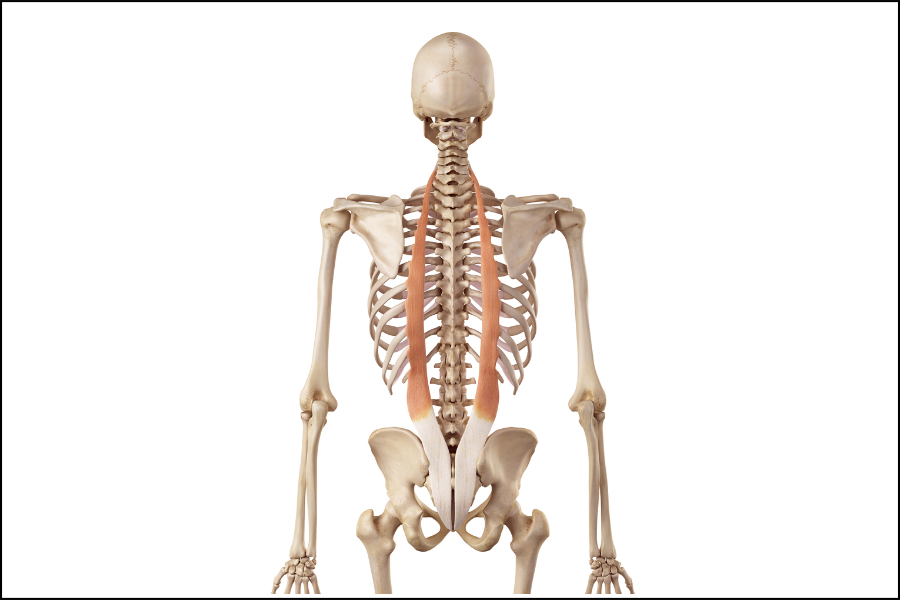
| Longissimus thoracis | Thoracolumbar fascia, transverse processes of lumbar vertebrae, and shared tendon with iliocostalis lumborum. | Transverse processes of T1-12, angle of ribs 7-12 | Bilateral contraction: extension of spine, depresses ribs Unilateral contraction: lateral flexion of spine | Spinal nerves |
| Multifidus | Transverse processes of C3-L5 vertebrae, sacrum, PSIS, and posterior sacroiliac ligament | Spinous processes 2-4 vertebral levels superior to their origin | Bilateral contraction: extension of spine Unilateral contraction: rotation of spine to the opposite (contralateral) side | Cervical, thoracic and lumbar spinal nerves |
| Quadratus lumborum | Posterior part of iliac crest, iliolumbar ligament | Transverse processes of L1-4 vertebrae, and inferior border of the 12th rib | Extension and lateral flexion of trunk. Elevation of ipsilateral pelvis. Depresses the 12th ribs. | Lumbar spinal nerves (L1-4); subcostal nerve. |
| Rhomboid minor | Spinous processes of vertebrae C7-T1, and inferior part of nuchal ligament | Medial (vertebral) border of scapula, at root of spine of scapula | Retraction and downward rotation of scapula | Dorsal scapular nerve (C5) |
| Rhomboid major | Spinous processes of vertebrae T2-T5 | Medial (vertebral) border of scapula, from root of spine to inferior angle of scapula | Retraction and downward rotation of scapula | Dorsal scapular nerve (C5) |
| Rotatores | Transverse processes of each vertebra | Rotatores brevis: spinous process of vertebra 1 level above origin Rotatores longus: spinous process of vertebra 2 levels above origin | Bilateral contraction: extension of spine Unilateral contraction: rotation of spine to the opposite side | Spinal nerves C1-L5 |
| Semispinalis | Transverse processes of C5-T10 | Capitis: between superior and inferior nuchal lines of the occipital bone Cervicis & thoracis: spinous processes of 4-6 vertebrae above origin | Extension and lateral flexion of the spine. Rotation of spine to the opposite (contralateral) side. | Spinal nerves (C1-T12) |
| Serratus posterior superior | Nuchal ligament and spinous processes of C7-T3 | Superior borders of ribs 2-5, just lateral to their angles | Pulls the upper ribs upward to expand the thoracic cavity and aid in respiration | Intercostal nerves T2-5 |
| Serratus posterior inferior | Thoracolumbar fascia and spinous processes of T11-L3 | Inferior borders of ribs 9-12, just lateral to their angles | Pulls the lower ribs downward and outward to counteract the pull of the diaphragm and aid in respiration | Intercostal nerves T9-12 |
| Spinalis capitis | Spinous processes of C7-T1 | Occipital bone | Bilateral contraction: extension of cervical spine Unilateral contraction: lateral flexion of cervical spine to same side | Cervical spinal nerves |
| Spinalis cervicis | Inferior part of nuchal ligament and spinous processes of C7-T1 | Spinous processes of C2-4 | Bilateral contraction: extension of cervical spine Unilateral contraction: lateral flexion of cervical spine to same side | Cervical spinal nerves |
| Spinalis thoracis | Spinous processes of T11-L2 | Spinous processes of T2-8 | Bilateral contraction: extension of thoracic spine Unilateral contraction: lateral flexion of thoracic spine to same side | Thoracic spinal nerves |
| Trapezius | External occipital protuberance, medial third of superior nuchal line, ligamentum nuchae, spinous processes of vertebrae C7-T12 | Lateral third of clavicle, acromion process and upper crest of the scapular spine | Elevation, depression, retraction or upward rotation of scapula (depending on which part of the muscle contracts). Cervical extension (upper trapezius). | Accessory nerve (CNXI), and cervical nerves (C3,4) |

Become an MBLExGuide member
Becoming a member of MBLExGuide gives you access to the best MBLEx test prep materials on the market. Practice realistic exams and quizzes. Review all content areas on the massage licensing exam in our MBLEx Prep Course. Learn muscles with our Muscle Flashcards.