Putting the cart before the… snake?

Time to reexamine some assumptions (again)! And also, talk about Hox genes, because do I even need a reason?

Hox genes often come up when we look for explanations for various innovations in animal body plans – the digits of land vertebrates, the limbless abdomens of insects, the various feeding and walking and swimming appendages of crustaceans, the strongly differentiated vertebral columns of mammals, and so on.

Speaking of differentiated vertebral columns, here’s one group I’d always thought of as having pretty much the exact opposite of them: snakes. Vertebral columns are patterned, among other things, by Hox genes. Boundaries between different types of vertebrae such as cervical (neck) and thoracic (the ones bearing the ribcage) correspond to boundaries of Hox gene expression in the embryo – e.g. the thoracic region in mammals begins where HoxC6 starts being expressed.

In mammals like us, and also in archosaurs (dinosaurs/birds, crocodiles and extinct relatives thereof), these boundaries can be really obvious and sharply defined – here’s Wikipedia’s crocodile skeleton for an example:

In contrast, the spine of a snake (example from Wikipedia below) just looks like a very long ribcage with a wee tail:

Snakes, of course, are rather weird vertebrates, and weird things make us sciencey types dig for an explanation.

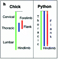
Since Hox genes appear to be responsible for the regionalisation of vertebral columns in mammals and archosaurs, it stands to reason that they’d also have something to do with the comparative lack of regionalisation (and the disappearance of limbs) seen in snakes and similar creatures. In a now classic paper, Cohn and Tickle (1999) observed that unlike in chicks, the Hox genes that normally define the neck and thoracic regions are kind of mashed together in embryonic pythons. Below is a simple schematic from the paper showing where three Hox genes are expressed along the body axis in these two animals. (Green is HoxB5, blue is C8, red is C6.)

Cohn_Tickle1999_hoxRegions

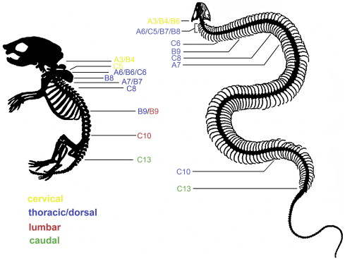
As more studies examined snake embryos, others came up with different ideas about the patterning of serpentine spines. Woltering et al. (2009) had a more in-depth look at Hox gene expression in both snakes and caecilians (limbless amphibians) and saw that there are in fact regions ruled by different Hoxes in these animals, if a little fuzzier than you’d expect in a mammal or bird – but they don’t appear to translate to different anatomical regions. Here’s their summary of their findings, showing the anteriormost limit of the activity of various Hox genes in a corn snake compared to a mouse:

Woltering_etal2009-mouse_vs_snake

Such differences aside, both of the above studies operated on the assumption that the vertebral column of snakes is “deregionalised” – i.e. that it evolved by losing well-defined anatomical regions present in its ancestors. But is that actually correct? Did snakes evolve from more regionalised ancestors, and did they then lose this regionalisation?

Head and Polly (2015) argue that the assumption of deregionalisation is a bit stinky. First, that super-long ribcage of snakes does in fact divide into several regions, and these regions respect the usual boundaries of Hox expression. Second, ordinary lizard-shaped lizards (from which snakes descended back in the days of the dinosaurs) are no more regionalised than snakes.

The study is mostly a statistical analysis of the shapes of vertebrae. Using an approach called geometric morphometrics, it turned these shapes from dozens of squamate (snake and lizard) species into sets of coordinates, which could then be compared to see how much they vary along the spine and whether the variation is smooth and continuous or clustered into different regions. The authors evaluated hypotheses regarding the number of distinct regions to see which one(s) best explained the observed variation. They also compared the squamates to alligators (representing archosaurs).

The results were partly what you’d expect. First, alligators showed much more overall variation in vertebral shape than squamates. Note that that’s all squamates – leggy lizards are nearly (though not quite) as uniform as their snake-like relatives. However, in all squamates, the best-fitting model of regionalisation was still one with either three or four distinct regions in front of the hips/cloaca, and in the majority, it was four, the same number as the alligator had.

Moreover, there appeared to be no strong support for an evolutionary pattern to the number of regions – specifically, none of the scenarios in which the origin of snake-like body plans involved the loss of one or more regions were particularly favoured by the data. There was also no systematic variation in the relative lengths of various regions; the idea that snakes in general have ridiculously long thoraxes is not supported by this analysis.

In summary, snakes might show a little less variation in vertebral shape than their closest relatives, but they certainly didn’t descend from alligator-style sharply regionalised ancestors, and they do still have regionalised spines.

Hox gene expression is not known for most of the creatures for which vertebral shapes were analysed, but such data do exist for mammals (mice, here), alligators, and corn snakes. What is known about different domains of Hox gene activation in these three animals turns out to match the anatomical boundaries defined by the models pretty well. In the mouse and alligator, Hox expression boundaries are sharp, and the borders of regions fall within one vertebra of them.

In the snake, the genetic and morphological boundaries are both gradual, but the boundaries estimated by the best model are always within the fuzzy boundary region of an appropriate Hox gene expression domain. Overall, the relationship between Hox genes and regions of the spine is pretty consistent in all three species.

To finish off, the authors make the important point that once you start turning to the fossil record and examining extinct relatives of mammals, or archosaurs, or squamates, or beasties close to the common ancestor of all three groups (collectively known as amniotes), you tend to find something less obviously regionalised than living mammals or archosaurs – check out this little figure from Head and Polly (2015) to see what they’re talking about:

Head_Polly2015-phylogeny_of_spines

(Moving across the tree, Seymouria is an early relative of amniotes but not quite an amniote; Captorhinus is similarly related to archosaurs and squamates, Uromastyx is the spiny-tailed lizard, Lichanura is a boa, Thrinaxodon is a close relative of mammals from the Triassic, and Mus, of course, is everyone’s favourite rodent. Note how alligators and mice really stand out with their ribless lower backs and suchlike.)

Although they don’t show stats for extinct creatures, Head and Polly argue that mammals and archosaurs, not snakes, are the weird ones when it comes to vertebral regionalisation. For most of amniote evolution, the norm was the more subtle version seen in living squamates. It was only during the origin of mammals and archosaurs that boundaries were sharpened and differences between regions magnified. Nice bit of convergent/parallel evolution there!

***

References:

Cohn MJ & Tickle C (1999) Developmental basis of limblessness and axial patterning in snakes. Nature 399:474-479

Head JJ & Polly PD (2015) Evolution of the snake body form reveals homoplasy in amniote Hox gene function. Nature 520:86-89

Woltering JM et al. (2009) Axial patterning in snakes and caecilians: evidence for an alternative interpretation of the Hox code. Developmental Biology 332:82-89
I couldn’t resist

Damn, I said I wasn’t going to talk about the Moroccan helicoplacoid-on-stalk, but it’s just so. Bloody. Amazing.

Here it is in its full glory, from the supplementary figures of Smith and Zamora (2013). Left is a cast of a young specimen, right is the authors’ reconstruction of the adult creature:

helicocystis_casthelicocystis_recon

So… the thing is a transitional form all right. It’s got a little stalk and cup like eocrinoids, built with a rather irregular arrangement of mineralised plates. On top of that it has a spiral body like helicoplacoids. It has ambulacra, the “rays” with porous plates where the tube feet that characterise living echinoderms can come out. This photo of the underside of a starfish is a pretty nice illustration of ambulacra (the white regions with little holes) and tube feet:

Even more interestingly, the new beastie (christened Helicocystis moroccoensis by the authors) seems to have five of them, like modern echinoderms (and a lot of extinct types, including eocrinoids). Helicoplacoids do have ambulacra, but only three or a single Y-shaped one, depending on interpetation.

Again unlike (one interpretation of) helicoplacoids but like modern echinoderms, the mouth of Helicocystis is right at the stalkless end. It’s also surrounded by an arrangement of skeletal plates that resembles more “conventional” echinoderms and has no equivalent in helicoplacoids proper. It’s about as neat a transitional form as you could hope for.

The question is which way the transition goes. It could be that the familiar five-rayed echinoderms are derived from a helicoplacoid-like ancestor, going through something like this guy. Or it could be that helicoplacoids are actually weird even for echinoderms, and their ancestors were more conventional stalked, five-armed beasties that lost their proper echinoderm shapes via something like Helicocystis.

Smith and Zamora actually did a phylogenetic analysis, but it’s not that helpful IMO. The tree in the paper is very pretty, and it says Helicocystis is the next branch after helicoplacoids on the path leading to “proper” echinoderms. The tree in the supplementary figures actually has measures of statistical support on it – which pretty confidently put Helicoplacus, Helicocystis, and a bunch of less weird echinoderms, together.

However, the relationships within that group are, shall we say, a little bit fluid. Granted, I come from a more sequency background and don’t often have to deal with morphology-based trees or parsimony as the method of analysis – but I’d definitely view a 56% bootstrap support with a big dose of scepticism, and this is the number they got for the hypothesis that Helicocystis is more closely related to “proper” echinoderms than to Helicoplacus. The other measure they display doesn’t make me any more confident about the relationship.

(I find it kind of amazing they got any resolution at all in that tree – with only 17 characters, some of which aren’t applicable to all species, and only nine species to begin with… yeah. The whole phylogenetic analysis is far from ideal even if it’s the best they could think of.)

So, based on that tree, the phylogenetic hypothesis they present is, at this point, just a plausible hypothesis. That doesn’t lessen the value of Helicocystis, though. The creature is still a damn neat transitional form – we just can’t be terribly sure which way the transition went.

There’s some interesting speculation in the paper about developmental evolution (yay!). Smith and Zamora point out that the spirally bit in Helicocystis looks like a complete helicoplacoid; the stalk and cup are kind of tacked onto that. The tissues of most modern echinoderm adults come from two different places: regular old tissues of the larva, and a special set of cells set aside for adult-making purposes*. So Smith and Zamora hypothesise that the two-part body of Helicocystis marks the point where this dual origin appeared. (Or, if they’re wrong about the phylogeny, the point where proto-helicoplacoids lost it?)

There’s also another interesting bit of evo-devo speculation (mixed with a bit of “eco”) about the stalk. Full-grown Helicocystis have pretty small stalks compared both to their own young and more typical stalked echinoderms. The authors wonder if this is because stalks for attachment originally functioned to help young echinoderms settle in a comfortable place, and only later became important for adults. I’m not sure how much sense that actually makes, and of course we only have a single species of Helicocystis to go by, but hey, ideas are fun.

Helicocystis has a random weird quirk as well, in that its spirals curl the opposite way to every proper helicoplacoid. That sort of variation happens even within species (e.g. in snail shells), but isn’t it a weird coincidence that such a unique creature should also twist the wrong way?

One thing is for sure: this beast is made of pure, distilled awesome. I think we should make a new Archaeopteryx out of it. Invertebrates need their evolutionary icons, too!

***

*And that’s a nice reminder for me, because I thought they basically threw away the larva. Apparently I need a refresher on echinoderm development. Or just a reminder that not all echinoderms are sea urchins. The funny thing is a couple of years ago I actually specifically read and puzzled over literature discussing what comes from where in various echinderms…

***

Reference:

Smith AB, Zamora S (2013) Cambrian spiral-plated echinoderms from Gondwana reveal the earliest pentaradial body plan. Proceedings of the Royal Society B advance online publication 26/06/2013, doi:10.1098/rspb.2013.1197

Echinoderm bonanza

Smith et al. (2013) has been sitting on my desktop waiting to be read for the last month or so. Man, am I glad that I finally opened the thing. I’m quite fond of echinoderms, and this paper is full of them. Of course. It’s about echinoderms. Specifically, it’s about the diverse menagerie of them that existed, it seems, a little bit earlier than thought.

The brief little paper introduces new echinoderm finds from two Mid-Cambrian formations in Morocco, which at the time was part of the great continent of Gondwana. As far as I’m concerned, it was worth reading just for this lineup of Cambrian echinoderms. I mean, echinoderms are so amazingly weird in such a variety of ways. They’re a delight.

Smith_etal2013-fig3_decorated

(The drawings themselves are from Fig. 3. of the paper; I rearranged them to fit into my post width, and the boxes are my additions. Dark box = new groups/species from Morocco, light grey box = known groups/species whose first appearance was pushed back in time by the Moroccan finds.)

Although none of the creatures above belong to the living classes of echinoderms, they display a wide range of body plans. You could say their body plans are more diverse* than those of living echinoderms. (And if you said that, the ghost of Stephen Jay Gould would nod approvingly.) For example, modern echinoderms tend to have either (usually five-part) radial symmetry (any old starfish) or bilateral symmetry that clearly comes from radial symmetry (heart urchins).

In these Early- to Mid-Cambrian varieties, you can see some five-rayed creatures, some that are more or less bilateral without any obvious connection to the prototypical five-point star, animals that are just kind of asymmetric, and those strange spindle-shaped helicoplacoids that look like someone took an animal with radial symmetry and wrung it out. And then there are all the various arrangements of arms and stalks and armour plates that I tend to gloss over when reading about the beasts. (Yeah. I have no attention span.)

The Morroccan finds have some very interesting highlights. The second creature in the lineup above is one of them. Its top half looks like a helicoplacoid such as Helicoplacus itself (first drawing). It’s got that characteristic spiral arrangement of plates and a mouth at the top end. However, unlike previously known helicoplacoids, it sits on a stalk that resembles the radially-symmetric eocrinoids (like the creature on its right). It’s a transitional form all right, though we’ll have to wait for future publications and perhaps future discoveries to see which way evolution actually went. It’ll already help palaeontologists make sense of helicoplacoids themselves, though, which I gather is a big thing in itself. The authors promise to publish a proper description of the creature, which is really exciting.

The other exciting thing about the Moroccan echinoderms is their age. As I already hinted at with my grey boxes, the new fossils push back the known time range of many echinoderm body plans by millions of years. This means that the wide variety of body plans we saw above was already present as little as 10-15 million years after the first appearance of scattered bits of echinoderm skeleton in the fossil record.

Smith et al. argue that this is a fairly solid conclusion based on the mineralogy of echinoderm skeletons. Organisms with calcium carbonate hard parts have a tendency to adopt the “easiest” mineralogy at the time they first evolve skeletons. Seawater composition changes over geological time; most importantly, the ratio of calcium to magnesium fluctuates. Calcium carbonate can adopt several different crystal forms, and the Ca/Mg ratio influences which of them are easier to make. So when there’s a lot of Mg in the sea, aragonite is the “natural” choice, whereas low Mg levels favour calcite.

The first appearance of echinoderms around 525 million years ago coincides with a shift in ocean chemistry from “aragonite seas” to “calcite seas”. Echinoderms and a bunch of other groups that first show up around that time have skeletons that are calcite in their structure but incorporate a lot of Mg. Since the ocean before was favourable to aragonite, it’s unlikely that echinoderm skeletons appeared much earlier than this date. In other words, echinoderm evolution during this geologically short period was truly worthy of the name “Cambrian explosion”.

That is, of course, if the appearance of echinoderm skeletons precedes the appearance of echinoderm body plans. The oldest of our Cambrian treasure troves of soft-bodied fossils, such as the rocks that yielded the Chengjiang biota of China, are roughly the same age as the first echinoderm skeletons. However, they don’t contain undisputed echinoderms as far as I can tell (Clausen et al., 2010). Proposed “echinoderms” from before the Cambrian are even less accepted. Of course, the unique structure of echinoderm skeletons is easy to recognise, but how do you identify an echinoderm ancestor without such a skeleton? (Is all that bodyplan diversity even possible without hard skeletal support?)

Caveats aside, this Moroccan stuff is awesome. And also, if my caveat proves overly cautious, echinoderms did some serious evolving in their first few million years on earth. A supersonic ride with Macroevolution Airlines?

***

*OK, if I want to be absolutely pedantic, and I do, then body plans are disparate rather than diverse. “Disparity” in palaeontological/evo-devo parlance refers to how different two or more creatures are. Diversity means how many different creatures there are. Maybe I should do a post on that, actually.

***

References:

Clausen S et al. (2010) The absence of echinoderms from the Lower Cambrian Chengjiang fauna of China: Palaeoecological and palaeogeographical implications. Palaeogeography, Palaeoclimatology, Palaeoecology 294:133-141

Smith AB et al. (2013) The oldest echinoderm faunas from Gondwana show that echinoderm body plan diversification was rapid. Nature Communications 4:1385

Another man after my own heart

It’s not terribly hard to turn me into a squealing fangirl. One of the ways is to agree with me eloquently and/or share my pet peeves. Another is to give me lightbulb moments. A third is to disagree with me in a well-reasoned, intelligent way. And finally, if I see you thoughtfully examining your own thinking, you are awesome by definition. Michaël Manuel’s monster review of body symmetry and polarity in animals (Manuel, 2009) did all of the above.

(In case you wondered, that means a long, squeeful meandering >.>)

Manuel writes about the evolution of two fundamental properties of animal body plans [1]: symmetry and polarity. You probably have a good intuitive understanding of symmetry, but here’s a definition anyway. An object is symmetrical if you can perform some transformation (rotation, reflection, shifting etc.) on it and get the same shape. Polarity is a different but equally simple concept – it basically means that one end of an object is different from the other, like the head and tail of a cat or the inner and outer arcs of a rainbow.

I can’t say that I’d thought an awful lot about either before I came across this review, so it’s not really surprising that I had lightbulbs going off in my head left and right while I was reading it. Because I didn’t think deeply about symmetry and polarity and complexity, I basically held the mainstream view I – and, I suspect, most of the mainstream – mostly picked up by osmosis.

That meant I fell victim to my own biggest pet peeve big time – I believed, without good reason and without even realising, that the body plan symmetries of major lineages of living animals represented successive increases in complexity. Sponges are kind of asymmetrical, cnidarians and ctenophores are radially symmetrical, and bilaterians such as ourselves have (more or less) mirror image symmetry, and these kinds of symmetry increase in complexity in this order. Only… they aren’t, and they don’t.

It turns out that this guy not only shares my pet peeve but uses it to demolish my long-held hidden assumptions. Double fangirl points!

Let there be light(bulbs)!

Problem number one with the traditional view – aside from ignoring that evolution ain’t a ladder – is that the distribution of symmetry types among animals is a little more complicated. Most importantly, most kinds of sponges are not asymmetrical. Most species may be, but that’s not the same thing. You see, most sponge species are demosponges, which make up only one of the four great divisions among sponges. Demosponges do have a tendency towards looking a bit amorphous, but the other three – calcareous sponges, glass sponges and homoscleromorphs – usually are some kind of symmetrical. All in all, the evidence points away from an asymmetrical animal ancestor. (Below: calcareous sponges being blatantly symmetrical, from Haeckel’s Kunstformen der Natur.)

The second problem is that my old view ignores at least one important kind of symmetry. Some “radially” symmetrical animals are actually closer to cylindrical symmetry. To understand the difference, imagine rotating a brick and a straight piece of pipe around their respective long axes. You can rotate the pipe as much or as little as you like, it’ll look exactly the same. In contrast, the only rotation that brings the brick back onto itself is turning it by 180° or multiples thereof. A pipe, with its infinitely many rotational symmetries, is cylindrically symmetrical, while the brick has a finite number of rotational symmetries [2], making it radially symmetrical.

Problem number three is that bilateral symmetry is actually no more complex than radial symmetry! What does “complexity” mean in this context? Manuel defines it as the number of coordinates required to specify any point in the animal’s body. In an animal with cylindrical symmetry, you only need a maximum of two: where along the main body axis and how far from the main body axis you are. Everything else is irrelevant, since these are the only axes along which the animal may be polarised. (Add any other polarity axis, and you’ve lost the cylindrical symmetry.)

Take a radially symmetrical creature, like a jellyfish. These also have a main rotational axis and an inside-outside axis of polarity. However, now the animal’s circumference is also divided up into regions, like slices in a cake. How does a skin cell around a baby jelly’s mouth know whether it’s to grow out into a tentacle or contribute to the space between tentacles? That is an extra instruction, an extra layer of complexity. We’re up to three. (Incidentally, here’s some jellyfish symmetry from Haeckel’s Kunstformen. [Here‘s photos of the real animal] A big cheat he may have been, but ol’ Ernst Haeckel certainly had an eye for beauty!)

And with that, jellies and their kin essentially catch up to the basic bilaterian plan. Because what do you need to specify a worm? You need a head-to-tail coordinate, you need a top-to-bottom one, and you need to say how far from the plane of symmetry you are. Still only three! Many bilaterians, including us, added a fourth coordinate by having different left and right sides, but that’s almost certainly not how we started when we split from the cnidarian lineage. (Below: radial symmetry doesn’t hold a monopoly on beauty! Three-striped flatworm [Pseudoceros tristriatus] by wildsingapore.)

Not only that, but Manuel argues that there’s very little evidence bilateral symmetry evolved from radial symmetry. By his reckoning, the most likely symmetry of the cnidarian-bilaterian common ancestor was cylindrical and not radial (more on this later, though). Thus the (mostly) radial cnidarians and the (mostly) bilateral bilaterians represent separate elaborations of a cylinder rather than stages in the same process.

There were a bunch more smaller lightbulb moments, but I’m already running long, so let’s get on to other things.

Respectful disagreement

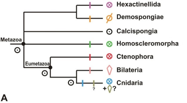
I think my disagreements with Manuel’s review are more of degree than of kind. Our fundamental difference of opinion comes back to the symmetries of various ancestors and the evidence for them. He argues that key ancestors in animal phylogeny – that of cnidarians + bilaterians, that of cnidarians + bilaterians + ctenophores, and that of all animals – were cylindrical. (Below is the reference tree Manuel uses for his discussion, with symmetry types indicated by the little icons.)

ManuelPhylo

I think he may well be correct in his conclusions, but I’m not entirely comfortable with his reasons. For example, he infers that the last common ancestor of cnidarians and ctenophores was cylindrical. One of his main arguments is that the repeated structures that “break up the cylinder” to confer radial symmetry are not the same in these two phyla. I think this is an intelligent point a smart guy who knows his zoology would make, so disagreement with it becomes debate as opposed to steamrolling [3].

Why I still disagree? As I said, it comes down to degrees and not kinds. Manuel considers the above evidence against a radially symmetrical common ancestor. I consider it lack of evidence for same. The situation reminds me of Erwin and Davidson (2002), which is also one of my favourite papers ever. They raise perhaps the most important point one could make about comparative developmental genetics: homologous pathways could have been present in common ancestors without the complex structures now generated by those pathways being there. Likewise, I think, radial symmetry could have been there in the common ancestor of cnidarians and ctenophores while none of the complex radially symmetrical structures (tentacles, stomach pouches, comb rows etc.) in the living animals were. Perhaps there were simpler divisions of cell types or whatnot that gave rise to the more overt radial symmetry of jellyfish, sea anemones and comb jellies.

In a related argument, Manuel discusses the homology (or lack thereof) of the dorsoventral axis in bilaterians and the so-called directive axis in sea anemones. Sea anemones actually show hints of bilateral symmetry, which prompted some authors (e.g. Baguñà et al., 2008) to argue that this bilateral symmetry and ours was inherited from a common ancestor (i.e. the cnidarian-bilaterian ancestor was bilateral).

I agree with Manuel that the developmental genetic evidence for this is equivocal at best. I even agree with him that developmental genetics isn’t decisive evidence for homology even if it matches better than it actually does in this case. But again, once the genetic evidence is dismissed as inconclusive, he relies on the non-homology of bilaterally symmetrical structures to conclude non-homology of bilateral symmetry. Again, I think this is a plausible but premature inference. Since I’m not sure whether homology or independent origin of bilateral symmetry is the better default hypothesis in this case, and I don’t think the evidence for/against either is convincing, I actually wouldn’t come down on either side as of yet.

But I can see his point, and that’s really cool.

Why else you’re awesome, Michaël Manuel…

Because you have a whole rant about “basal lineages”. I grinned like a maniac throughout your penultimate paragraph. Incidentally, you might have given me another favourite paper – anything with “basal baloney” in its title sounds like it’s worth a few squees of its own!

Because you apply critical thinking to your own thinking. See where we disagreed, non-homology of structures vs. symmetries, evidence against vs no evidence for, and all that? After you made the argument from non-homology of structures, I expected you to leave it at that. And you didn’t. You went and acknowledged its limitations, even though you stood by your original conclusions in the end.

Because you reminded me that radial symmetry is similar to metamerism/segmentation. I’d thought of that before, but it sort of went on holiday for a long time. Connections, yay!

Because you were suspicious about sponges’ lack of Hox/ParaHox genes. And how right you were!

*

Phew, that turned out rather longer and less coherent than I intended. And I didn’t even cover half of the stuff in my notes. I obviously really, really loved this paper…

***

[1] Or any body plan, really…

[2] Astute readers might have noticed that a brick has more than one axis of symmetry, plus several planes of symmetry as well. So it’s not only radially but also bilaterally symmetrical. The one thing it certainly isn’t is cylindrical 😉

[3] Not to say I don’t enjoy steamrolling obvious nonsense, but I also like growing intellectually, and steamrolling obvious nonsense rarely stretches the mind muscles…

***

References:

Baguñà J et al. (2008) Back in time: a new systematic proposal for the Bilateria. Philosophical Transactions of the Royal Society B 363:1481-1491

Erwin DH & Davidson EH (2002) The last common bilaterian ancestor. Development 129:3021-3032

Manuel M (2009) Early evolution of symmetry and polarity in metazoan body plans. Comptes Rendus Biologies 332:184-209

Neurulating acorn worms, and the Mammal is confused

To the everyday animal lover, acorn worms have few things going for them. They are pretty hideous as a rule, and they don’t really do anything interesting. (If you count “eating mud all day” as interesting, you are probably a sediment ecologist…)

True to their mud-eating selves, though, acorn worms are great at muddying the water in the evo-devo world. You see, there’s this neat idea that goes back to the early 19th century called dorsoventral inversion. French naturalist Étienne Geoffroy Saint-Hilaire once noted that an arthropod looks rather like an upside down vertebrate in many ways. This diagram from the Wikipedia entry illustrates the concept rather nicely:

The pink and snot-green triangles on the side represent the expression levels of the key genes that determine where the back and belly sides go. dpp (decapentaplegic) is the fly homologue of BMP4 (or BMP2, 4 and 7, to be precise), and sog (short gastrulation) is the fly version of chordin. So not only do major structures like the central nervous system develop on opposite sides of the body, the developmental genetics are also upside down. It seems like a no-brainer. (Well, chordates had to somehow move the mouth back to the belly side, but hey, no big deal ;))

Then those evil acorn worms come in and complicate things.

Acorn worms belong to the phylum Hemichordata. Hemichordates are most closely related to echinoderms like sea urchins. Together with chordates (vertebrates, sea squirts and lancelets), they make up the deuterostomes. Fruit flies and most worms that aren’t “acorn”, on the other hand, are protostomes.

Whether acorn worms and other hemichordates have a central nervous system at all has been the subject of some debate. Recent evidence suggests that they do (Nomaksteinsky et al., 2009). They don’t even just have one, they have two of them, taking the form of nerve cords on the dorsal and ventral sides of the animal. The dorsal nerve cord forms in a way eerily similar to our own spinal cord. Chordates like ourselves undergo a process called neurulation, in which some of the ectoderm (“skin”) on the embryo’s back folds inwards to form a hollow tube that’s the precursor of the central nervous system. Well, look what happens in an acorn worm (figure from Luttrell et al., 2012):

The left side of this image depicts the process in the acorn worm Ptychodera flava (and shows you a young worm – innit cute?), while the right side illustrates what happens in a chick embryo. (I’m not sure why they didn’t use actual photos of chick neurulation, I don’t imagine they’d be very hard to come by…)

In chordates, the notochord (precursor of our spine) sends out chemical signals to direct nerve cord formation. Acorn worms don’t have anything notochord-like at the time the nerve cord develops, but somehow they make it look like neurulation anyway. If it looks like a duck (or chick, as the case may be) and quacks like one, it might just be one.

The problem?

It happens on the wrong side. Having read the two papers I’ve cited so far, my impression is that neurulation only happens with the dorsal cord. However, in other respects these animals share the arthropod, not the vertebrate, orientation. Most importantly, their genetic dorsoventral axis is oriented like that of arthropods and other protostomes, with BMP levels in the embryo highest on the dorsal side (Lowe et al., 2006). Chordate embryos can’t even make a nervous system at high BMP levels!

It seems that whatever happened to turn the ancestral bilaterian on its head, it wasn’t as simple as flipping the animal and relocating the mouth. The development of the central nervous system shares serious developmental genetic similarities among deuterostomes like us and protostomes like flies (Arendt and Nübler-Jung, 1999) or ragworms (Denes et al., 2007), indicating that if not a full-blown CNS, then at least the genetic pattern was present in our common ancestor.

The figure above is a schematic comparison of gene expression in the developing nervous systems of fruit flies (Drosophila), ragworms (Platynereis) and vertebrates from Denes et al. (2007). The diagram labelled Enteropneust illustrates the lack of data from acorn worms, which I don’t fully understand given that Lowe et al. (2006) actually studied several of the genes included in this figure. Speaking of that, acorn worms once again prove to be weird and confusing, since the genes in question aren’t expressed in anything like the longitudinal stripes seen in the other animals. In fact, Lowe et al. (2006) found that they aren’t obviously associated with either of the nerve cords.

To be precise, Lowe et al. worked under the assumption that acorn worms had no nerve cords, but if you look at their pictures the lack of resemblance is blindingly obvious. For example, Msx, the light grey gene on the Platynereis and vertebrate diagrams, goes all around acorn worm embryos in a relatively narrow ring. Nk2.2, the red gene, isn’t even expressed in the ectoderm (the embryonic “skin”), whereas central nervous systems invariably come from there. Did Lowe et al. get the wrong genes or what? Don’t think so, Msxes at least are pretty easy to recognise…

To summarise: acorn worm central nervous systems develop much like ours, but on the wrong side of the body, with none of the genetic similarities we share with animals much less closely related to us than acorn worms. To top that, the damn worms have another nerve cord on the opposite side, which doesn’t develop by neurulation unless I’ve misunderstood something.

I… have no idea what’s going on here. Damn you, nature, you need to work on your clarity.

***

References:

Arendt D & Nübler-Jung K (1999) Comparison of early nerve cord development in insects and vertebrates. Development 126:2309-2325

Denes AS et al. (2007) Molecular architecture of annelid nerve cord supports common origin of nervous system centralization in Bilateria. Cell 129:277-288

Lowe CJ et al. (2006) Dorsoventral patterning in hemichordates: insights into early chordate evolution. PLoS Biology 4:e291

Luttrell S et al. (2012) Ptychoderid hemichordate neurulation without a notochord. Integrative and Comparative Biology 52:829-834

Nomaksteinsky M et al. (2009) Centrarlization of the deuterostome nervous system predates chordates. Current Biology 19:1264-1269

The origin of Hox genes: a telltale neighbourhood

Gods, it’s been so hard to keep my mouth shut about this. A friend of mine just published a paper about Hox genes, and I’ve known about it for a while and it’s been keeping me crazy excited because it’s fascinating and, well: Hox genes! Now that it’s finally out, I can blather about it to my heart’s content, and so I will. Be prepared for a long ride 😉

First of all, a quick rundown of Hox genes for those who aren’t evo-devo geeks. These genes encode transcription factors – proteins that switch genes on/off. They are members of the large and distinguished class of homeobox genes, many of which play important roles in orchestrating embryonic development. Hox genes in particular are famous for laying out the plan for the head to tail axes of bilaterian animals, and for often sitting in neat clusters in the genome and being expressed along the body axis in the same order they are in the cluster. (Below: one of my favourite scientific figures ever, a fruit fly embryo stained in different colours for each of its Hox genes*. From Lemons and McGinnis [2006] via Pharyngula) In short, Hox genes are fucking awesome and extremely important to boot.

Tracing origins

One of the unresolved questions about Hox genes is exactly where they come from, and the new study draws some interesting conclusions regarding their origins. Before we delve into Mendivil Ramos et al. ( 2012) itself, perhaps it’s best to pull out my old sketch of animal phylogeny, because the relationships of the great old animal lineages are kind of important for the discussion. So this is the family tree of animals at first approximation (photos were all sourced from Wikimedia Commons; more info about them in my Nectocaris post):

Mendivil Ramos et al. follow one of the more popular resolutions of the question marks, in which cnidarians are closest to bilaterians and placozoans are the sister group to cnidarians+bilaterians. They don’t talk too much about ctenophores, but I’ll return to that later 🙂

Bilaterians all have Hox genes, and in most of them they do what they were originally discovered doing in fruit flies: patterning the anterior-posterior axis as they say in Jargonese. Some bilaterians have duplicated individual genes or even whole Hox clusters (we have four clusters, and salmon have as many as 13), but it’s pretty uncontroversial that a neat Hox cluster with representatives of most existing types of Hox genes was present already on the left side of the bilaterian box. So was the little sister of the Hox cluster, unimaginatively called the ParaHox cluster, which only contains three kinds of genes but operates in a similar way to its more famous sister (Brooke et al., 1998).

Where did Hox and ParaHox genes come from? Given the phylogeny of the genes, it’s likely that there was originally a small (maybe 2-3 genes) ProtoHox cluster that duplicated to give rise to both Hoxes and ParaHoxes. We know that cnidarians like sea anemones have both Hox and ParaHox genes, which behave somewhat like their bilaterian counterparts (Ryan et al., 2007). Therefore, the ProtoHox cluster must have existed before the common ancestor of these two great lineages.

Enter the Blob

What about placozoans? That’s where things get a bit complicated. Trichoplax, the mysterious little blob that is the only living representative of this oddball phylum, has only one Hox-like gene noncommittally named Trox-2. A relic of the ProtoHox era? Not really – in phylogenetic analyses of the protein sequence, it tends to group with the ParaHox gene Gsx, whereas you would expect a leftover ProtoHox gene to remain outside the Hox+ProtoHox clique.

Is Trox-2 a ProtoHox gene anyway? That would mean something weird happened in the evolution of Hox and ParaHox genes after the cluster duplication: Gsx (and its sisters Hox1-2) would have stagnated somewhere near its ancestral condition while all the other genes sped ahead. It’s a long shot, but evolution has been known to do strange things to gene sequences. Also, homeobox genes are often difficult to classify by sequence alone. Scientists typically use the DNA-binding region that the homeobox encodes for this purpose, but a homeodomain is only 60 amino acids and simply doesn’t contain enough information to place some problematic sequences. And unless we’re examining very closely related genes, the rest of the protein sequence is too different to be compared.

Guilt by association

However, there is another way of solving the mystery. Hox and ParaHox genes are not alone in the genome. They sit on huge chromosomes, and while they tend to banish non-*Hox genes from among them, the flanks of each cluster are populated by a variety of unrelated genes. The key thing is that Hox clusters and ParaHox clusters have different neighbours. Thus, looking at a problem gene’s neighbours can tell us what it is!

(Above: the neighbours of Trox-2. Yellow genes are ParaHox neighbours in humans, green genes are Hox neighbours, grey genes have no human counterparts, and orange genes are parts of both Hox and ParaHox neighbourhoods. From Mendivil Ramos et al. [2012])

This is exactly what happened. My lovely friend Olivia looked at the chunk of genomic sequence that contains Trox-2 and found about two dozen genes on it that had clear homologues in humans. She then tallied where each of the human homologues were, and behold: many of them crowded around ParaHox clusters (we also have several of those, courtesy of whole genome duplications), while only one was a Hox neighbour in humans. If Trox-2 were a ProtoHox, we’d expect a mixture of Hox and ParaHox neighbours, but that’s not what we find at all. Statistically speaking, it’s a no-brainer. Trox-2 is exactly where a ParaHox gene should be.

Ghosts in the genome

Now, we have a problem. If Trox-2 is a ParaHox gene, it must have come after the Hox/ParaHox duplication. So where the hell is the Hox cluster? Well, seeing as Trichoplax only has one ParaHox gene instead of the more typical three or so, gene loss certainly sounds like a possibility. Is there an “empty” Hox cluster lurking somewhere in the blob’s genome? Here, cnidarians turn out to be pretty helpful. After sequencing the genome of the sea anemone Nematostella vectensis, Putnam et al. (2007) attempted to reconstruct parts of the original chromosomes of the cnidarian-bilaterian ancestor. They called the results Putative Ancestral Linkage Groups, in other words, groups of genes that have stayed together since cnidarians and bilaterians diverged 600 or so million years ago.

One of these PALs contains over 200 conserved Hox neighbours, nearly all of which are present in Trichoplax. Strikingly, about half of them are close enough to one another that they are in the same chunk of sequence even though the Trichoplax genome hasn’t been stitched together to the level of whole chromosomes. That’s much more than you’d expect by chance. Trichoplax has a Hox locus without Hox genes, what Mendivil Ramos et al. call a ghost Hox locus.

Hox genes all the way down?

If you followed so far, you might have noticed that we’ve been pushing that elusive ProtoHox further and further back in animal evolution. It preceded bilaterians, it preceded cnidarians and bilaterians, and now it turns out it also preceded our split from placozoans. Will we find it if we look in the remaining animal lineages? Since a ctenophore genome hasn’t yet been released to the public, that question transforms into: will we find it in sponges?

The sponge Amphimedon queenslandica does have a publicly available genome, and much has been made of its apparent lack of many developmentally important transcription factor families (e.g. Larroux et al., 2008). It doesn’t have anything that looks like a Hox, ParaHox or ProtoHox gene, but what about the neighbourhoods?

Like that of Trichoplax, the Amphimedon genome sequence is in relatively small pieces, so a little clever statisticking was needed to decide whether it contains Hox, ParaHox or ProtoHox neighbourhoods. The starting points were the PAL of Hox neighbours mentioned above, and a PAL of ParaHox neighbours the team constructed using the human and Trichoplax genomes. These genes were distributed among many genomic scaffolds, but of course lacking chromosome-level information the group didn’t know whether any of these scaffolds are actually linked to each other in the sponge genome.

The solution was a simulation: take the number of genes in the PAL, take the number and size (in number of genes) of the thousands of Amphimedon scaffolds, and scatter the PAL members randomly among the scaffolds with the larger scaffolds proportionately more likely to receive a PAL gene. When all the PAL members are handed out, count the number of scaffolds with PAL members on them. Repeat this a thousand times, and you get an idea what the distribution of Hox and ParaHox neighbours would be if they weren’t clustered together. This approach showed that the real distribution is anything but random. Hox and ParaHox neighbours are clearly clustered in the sponge genome, and what’s more, they are clustered separately.

Still no ProtoHox locus, in other words. At some point in the murky depths of their ancestry, sponges lost bona fide Hox and ParaHox genes!

So…

That raises a couple of issues. First, where is the ProtoHox? Hox-like genes have never been found outside animals. These are smart people we’re talking about, so they checked the genome of the closest non-animal relative we have today, a choanoflagellate. Neither Hox/ParaHox nor ProtoHox neighbourhoods were there – the PAL genes didn’t cluster together any more than they would by chance. The whole *Hox phenomenon seems unique to animals (or else the choanoflagellate genome is totally scrambled). It appears that somewhere in our ancestry, ProtoHox gene(s) appeared and parted ways before sponges split from the rest of the animals. Since we have no surviving descendants of these ancestors outside of sponges and the rest of the animals, we’ll probably never find unduplicated descendants of the ProtoHox cluster.

Second, what happened in ctenophores? Everything we know about their genomes suggests that they completely lack Hox-like genes. Although there have been studies that placed them even further out than sponges (Dunn et al., 2008), it’s more likely that they are much closer to bilaterians than that (Philippe et al., 2011). I think I’m not the only one itching to examine a ctenophore genome for Hox neighbours…

And finally, if some distant ancestor of all animals had full-blown Hox and ParaHox clusters, what the heck was it doing with them? Was it something unexpectedly complex that would need genes for axial patterning? Are sponges and placozoans grossly simplified descendants of a much more complex ancestor, or did Hox-like genes only become involved in dividing up body axes later in evolution?

The more we learn the less we know. One thing is (once again) clear: assuming that a simple animal is a good proxy for an ancestral animal is a dangerous, dangerous assumption to make.

***

*Technically, fruit flies have twelve Hox genes, but only seven are shown in the image. Hox2/proboscipedia is a normal Hox gene involved in the development of mouthparts among others, but four more genes have completely lost their “canonical” Hox gene-like activities. That includes all three of Drosophila‘s weird triplicated Hox3 genes.

***

References

Brooke NM et al. (1998) The ParaHox gene cluster is an evolutionary sister of the Hox gene cluster. Nature 392:920-922

Dunn CW et al. (2008) Broad phylogenomic sampling improves resolution of the animal tree of life. Nature 457:745-759

Larroux C et al. (2008) Genesis and expansion of metazoan transcription factor gene classes. Molecular Biology and Evolution 25:980-996

Lemons D and McGinnis W (2006) Genomic evolution of Hox gene clusters. Science 313:1918-1922

Mendivil Ramos O et al. (2012) Ghost loci imply Hox and ParaHox existence in the last common ancestor of animals. Current Biology in press, available online 26/09/2012, doi: 10.1016/j.cub.2012.08.023

Philippe H et al. (2011) Resolving difficult phylogenetic questions: why more sequences are not enough. PLoS Biology 9:e1000602

Putnam NH et al. (2007) Sea anemone genome reveals ancestral eumetazoan gene repertoire and genomic organization. Science 317:86-94

Ryan JF et al. (2007) Pre-bilaterian origins of the Hox cluster and the Hox code: evidence from the sea anemone, Nematostella vectensis. PLoS ONE 2:e153